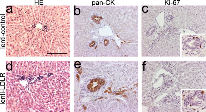
Figure 4.
Immunohistochemical characterization of representative rabbit liver samples after gene transfer with lentiviral vectors. Representative images of HE-stained liver 1 year after LV-LDLR (a) and LV-control (d) gene transfers. (b,e) Immunohistochemical staining with a pan-cytokeratin antibody stained large and small bile ducts in the liver in both groups. (c,f) Individual Ki-67 positive cells were seen in bile ducts of LV transduced animals one year after gene transfer. No significant difference was seen between the treatment groups. Magnification 200x, inserts 400x. Scale bar: 100 µm.

