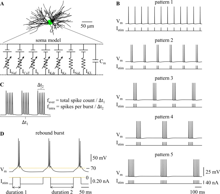
Figure 1.
Firing patterns in a computational model of a TC relay neuron. (A) Schematic of the model. (B) Temporal patterns of neural activity. Transmembrane voltage Vm was measured in the cell body. Depolarizing pulse train Istim (pulse width: 0.1 ms, pulse amplitude: 40 nA) was applied to cell body. (C) Graphical depiction of faver and fintra. faver = total spike count/Δt1, and fintra = spikes per burst/Δt2. (D) Rebound burst at faver = 10 Hz. Hyperpolarizing current (amplitude: −0.2 nA) was injected in the cell body. Duration 1 was 50 ms, and duration 2 was 150 ms.

