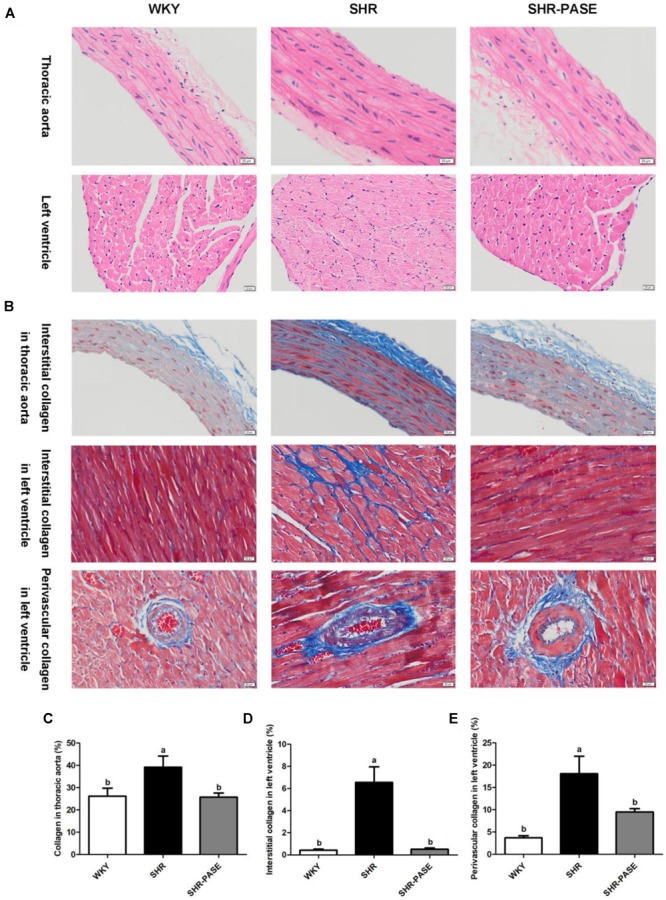
FIGURE 4.

Effect of PASE on target organ hypertrophy and fibrosis. Histology of thoracic aortal and left ventricular tissues from SHR after 12-week administration of PASE. H&E staining (A) were used to investigate the morphological changes and Masson staining (B) to examine fibrosis levels in the target organs (original magnification × 200). Contents of collagen in thoracic aorta (C), myocardial interstitial (D), and perivascular spaces (E) of left ventricle were also measured. Data are presented by means ± SE; n = 6 per group. Statistical analyses were performed by one-way ANOVA with Bonferroni post hoc test. Labeled means without a common letter indicated a significant difference at p < 0.05 (a > b) between groups.
