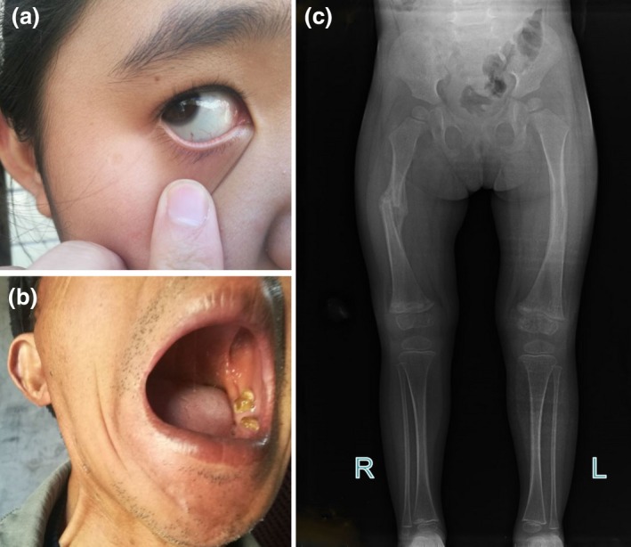
Figure 2.

(a) The proband (IV:1) presents with blue sclera. (b) Clinical picture shows dentinogenesis imperfecta in patient (II:2). (c) Radiograph shows fractures and abnormal callus formation of the proband (IV:1) resulting in slight deformations of long bones
