Figure 1.

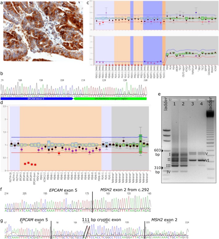
Representative molecular characterization of patient CFS396 carrying the EPCAM Del_16.5. (a) Immunohistochemistry showing that MSH2 protein is exclusively expressed in the cytoplasm of the tumor cells (H&E counterstain, O.M. 400x). (b) Sanger sequence of breakpoint. (c) Coffalyser analysis of methylation specific‐MLPA in tumor DNA, displaying MSH2 promoter methylation. (d) Coffalyser analysis of MLPA in tumor DNA, in which partial loss of heterozygosity of EPCAM is evidenced, due to a large somatic deletion involving also MSH2 and MSH6 genes. (e) Agarose‐gel electrophoresis showing aberrant EPCAM/MSH2 fusion transcripts amplified from tumor (lane 1–2) and normal mucosa (lanes 3–4) cDNAs; DNA ladders: ΦX174 DNA‐Hae III Digest (left) and 100 bp ladder (right). Roman numerals indicate the main PCR products that were sequenced. (f) Sanger sequence of an in‐frame fusion transcript (corresponding to amplicon III). (g) Sanger sequence of an out‐of‐frame fusion transcript (amplicon I and V)
