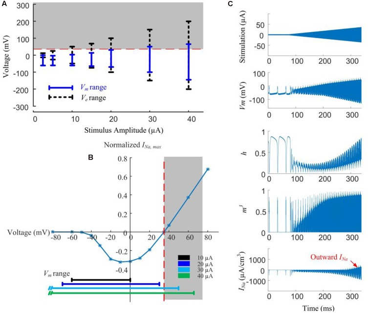
FIGURE 5.
Modeled reversal of sodium current with high-amplitude HFS induces spike inhibition. (A) Range of extracellular voltage (Ve) and transmembrane potential (Vm) determined from voltage value differences at cathodal and anodal phase peaks. The shaded region indicates the voltage range (>35 mV) where reversal of sodium current occurs. (B) Normalized I-V curve of model RGC peak sodium current. Peak sodium current (INa,max) becomes outward when Vm is above its reversal potential of 35 mV (the shaded region). This reversal occurs when the stimulus amplitude is higher than 20 μA. (C) Reversal of sodium current during 2-kHz amplitude-modulated stimulation (250 ms duration, 2 μA baseline ramping to a peak of 40 μA). For HFS amplitudes higher than 30 μA, the activation (m) and inactivation (h) gating variables of the sodium current (neurites below the electrode) are entrained by the extracellular voltage changes. The sodium current becomes increasingly outward with higher HFS amplitudes (indicated in the bottom right panel).

