Figure 1.
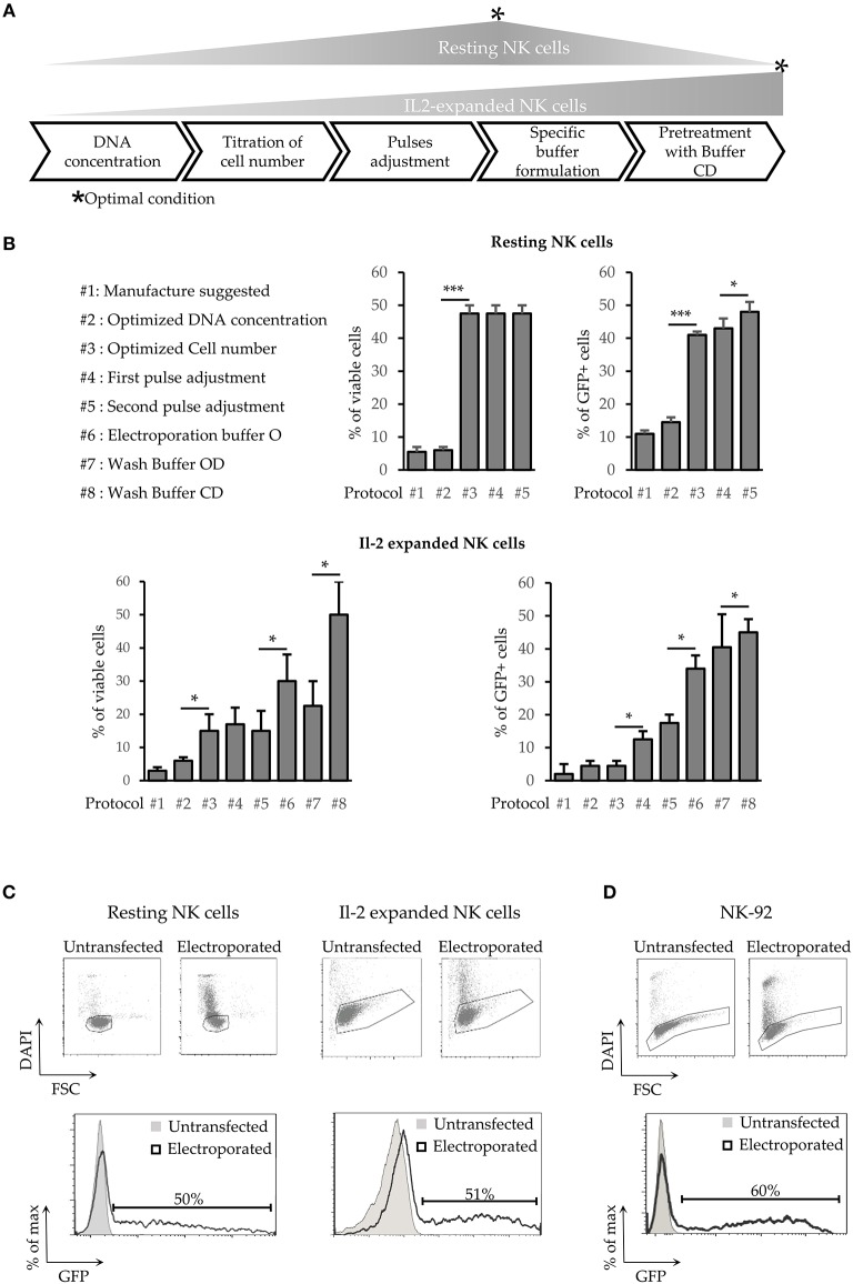
Development of a new transfection method for human NK cells. (A) Schematic steps of NK cell electroporation-based transfection methods. The asterisks (*) indicate the step in which we obtained the higher efficiency and viability (Optimal Condition O.C.) of resting or activated NK cells. (B) Percentages of cell viability and transfection efficiency obtained for the different protocols (from #1 to #8) applied to improve NK cell electroporation. Error bars indicate Standard Deviation (SD). P-values were calculated comparing each protocol with the previous one. *p < 0.05, **p < 0.01, and ***p < 0.001. (C,D) GFP expression in electroporated resting NK cells (C, left panel), Il-2 activated NK cells (C, right panel) and in NK92 cell line (D). One representative experiment out of 4 performed is shown.

