Figure 2.
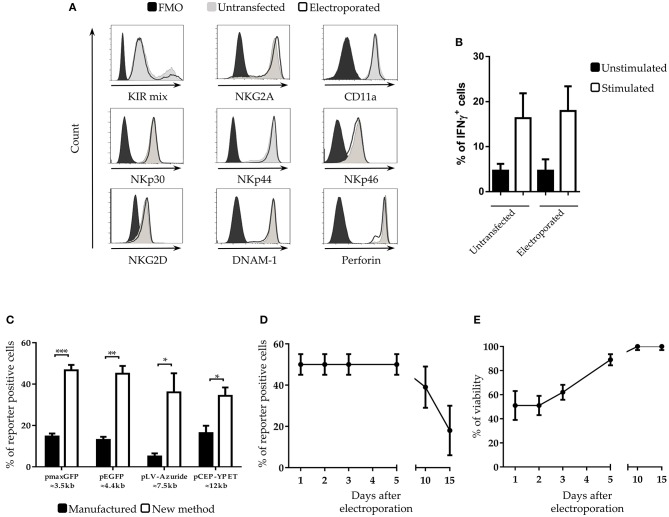
Characterization of NK cells after electroporation (A) Cytofluorimetric analysis of NK cell receptors and perforin expression on IL-2-activated NK cells at 5 days after electroporation in FMO control (black filled profiles), untrasfected (gray filled profiles) or with pmaxGFP plasmid (black empty profiles). A representative experiment out of 3 is shown. (B) Percentages of interferon-gamma (IFNγ) positive untransfected- and pmaxGFP electroporated-NK cells. Unstimulated (black bars) are compared with K562 stimulated (white bars) NK cells. (C) Transfection with different plasmid sizes in NK cells. Percentage of viability and efficiency after electroporation of activated NK cells with plasmids of different size. ***p < 0.001, **p < 0.01 and *p < 0.05. (D) Persistence of transfected genes and (E) viability of NK cells at different culture intervals after electroporation. Six experiments performed.

