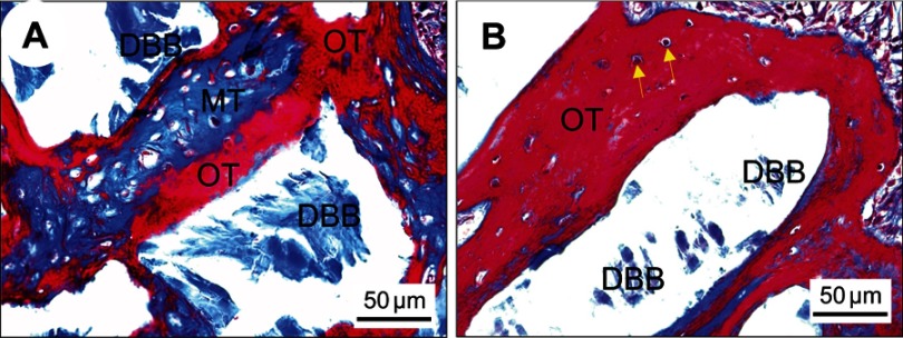
Figure 5.
Histological observation of bone restoration after 12 weeks post-implantation. Representative Masson’s trichrome staining images of DBB granules within defects covered with PTFE membranes (A) or BTO/P(VDF-TrFE) nanocomposite membranes (B). Yellow arrows denote viable osteocytes in their lacunae. Scale bar =50 μm.
Abbreviations: DBB, deproteinized bovine bone; OT, osteoid tissue (red); MT, mineralized tissue (blue); PTFE, polytetrafluoroethylene; BTO, BaTiO3; P(VDF-TrFE), poly(vinylidene fluoridetrifluoroethylene).

