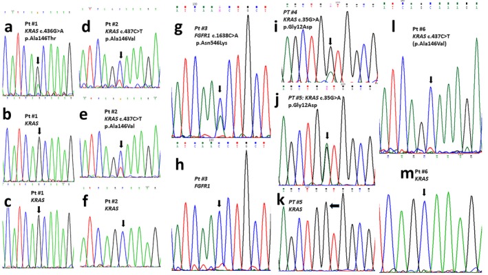
Figure 2.

Molecular analysis of RAS‐related genes. Patient #1 (a–c). (a) KRAS partial sequence showing the c.436G>A (p.Ala146Thr) mutation in DNA from the aplasia cutis lesion; This variant was not identified in DNA from buccal mucosa (b) or peripheral leukocytes (c). Patient #2 (d–f). (d,e). KRAS partial nucleotidic sequence showing c.437C>T (p.Ala146Val) mutation in DNA obtained from the aplasia cutis (d) and hyperpigmented skin (e) lesions. This variant was not identified in DNA from buccal mucosa or leukocytes (f). Patient #3 FGFR1 partial nucleotidic sequence showing the pathogenic c.1638C>A (p.Asn546Lys) variant identified in DNA from nevus psiloliparus (g). This variant was not identified in DNA from buccal mucosa or peripheral leukocytes (h). Patient #4 (i), patient #5 (j). KRAS partial DNA sequence showing the pathogenic variant c.35G>A (p.Gly12Asp) in DNA from nevus sebaceous of parietal region (i), and upper lip (j). This variant was not identified in DNA from buccal mucosa or blood leukocytes (k, only patient #5 sequence is shown)). Patient #6 (l, m). KRAS partial nucleotide sequence showing the c.437C>T (p.Ala146Val) mutation (l) identified in DNA from an epibulbar dermoid. This variant was not identified in DNA from buccal mucosa (m) or blood leukocytes
