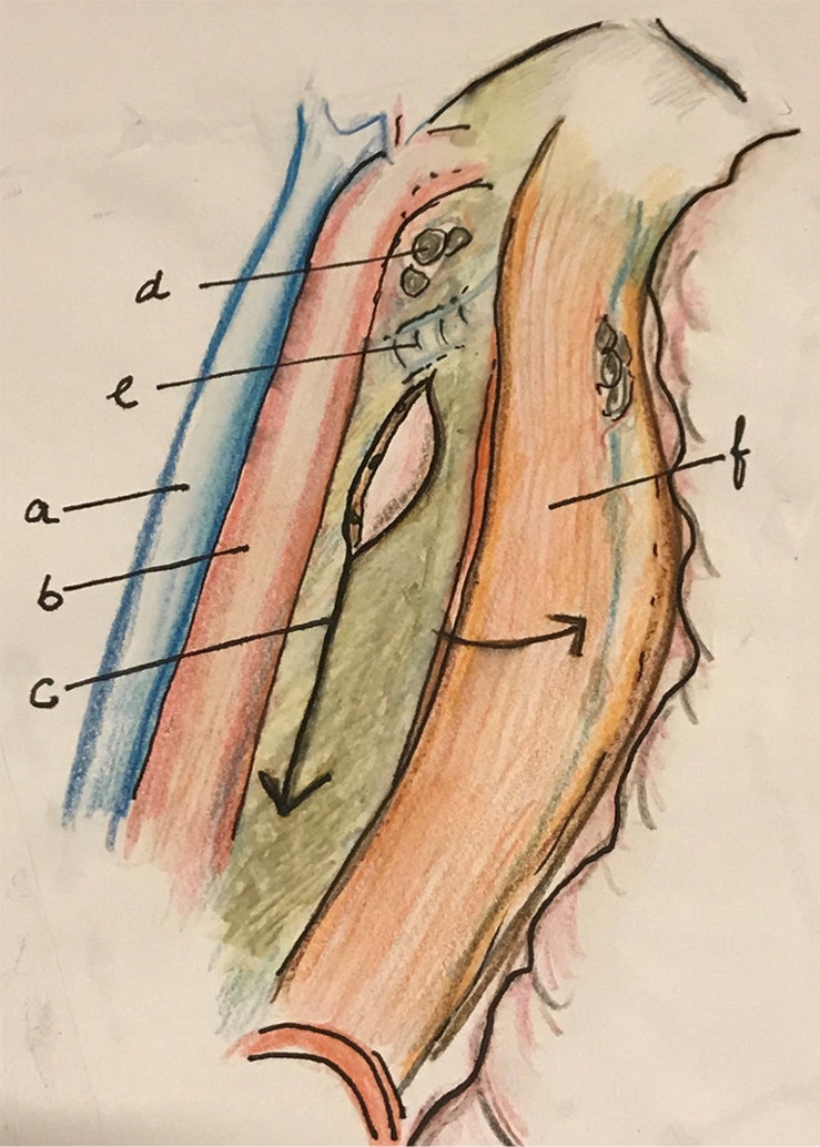
Figure 6.

After dissection along the descending aorta and dissection and resection of the thoracic duct, the esophagus is retracted to right to expose and divide the lower mesoesophagus. a, azygos vein; b, aorta; c, mesoesophagus (distal); d, subaortic LN; e, left bronchus; f, esophagus. LN, lymph nodes.
