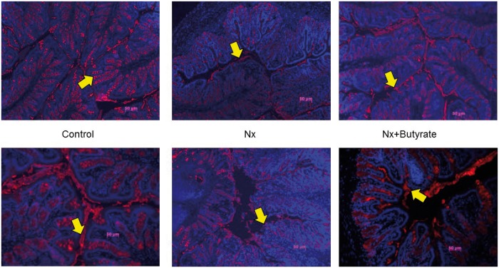
FIGURE 6.
Muc2 expression in the colon analyzed immunohistochemically revealed decreased expression of Muc2 in the Nx group was abrogated by butyrate. Fluorescent microscopy of Muc2, stained red, merged with DNA-capturing 4′-6-diamidino-2-phenylindole, stained blue. Representative sections of colonic mucin of two animals from each group (control, Nx and butyrate-treated Nx) are shown. A total of six animals per group were used to assess Muc2 expression. The proximal part of the colon (0.5 cm) was collected and stored in Carnoy’s solution. Muc2 expression was determined by immunohistochemistry using Muc2 antibody. The yellow arrow points to the Muc2 (red) expression. Compared with controls, Muc2 expression was significantly lower in the Nx groups. The butyrate-treated group had a significant increase in mucin expression.

