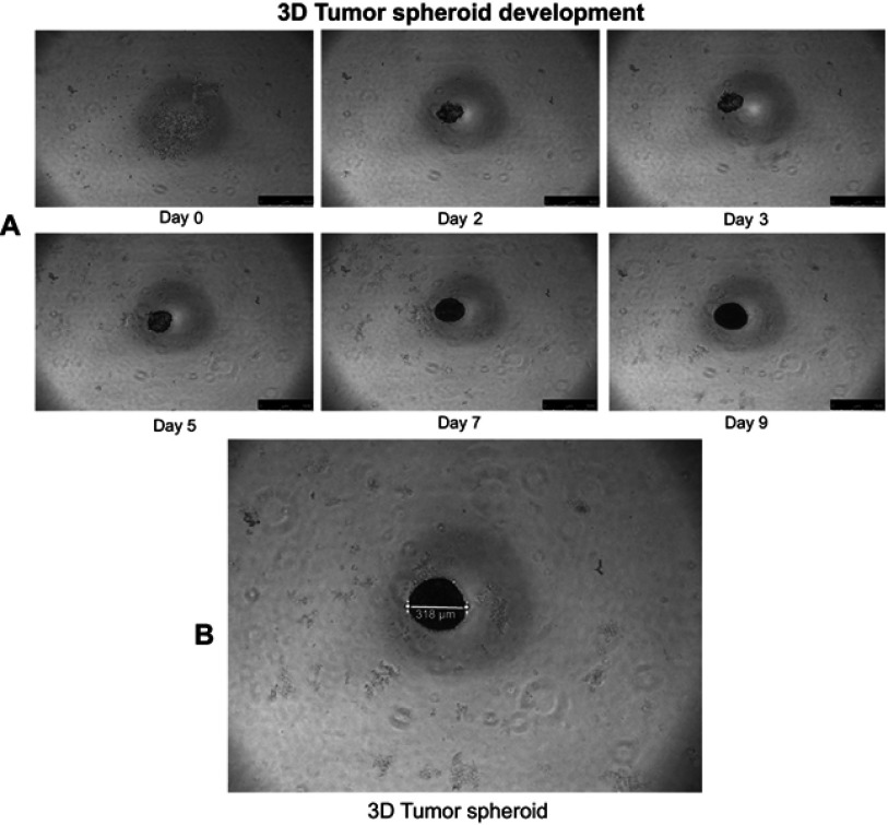
Figure S2.
Representative bright-field images of A549 tumor-spheroid development. (A) Development of three-dimensional (3D) lung tumor spheroid at different time points (0, 2, 3, 5, 7 and 9 days). (B) Compact and uniform tumor spheroid (>300 µm) were selected for use in tumor-spheroid penetration and growth inhibition studies. The scale bar represents 500 µm. The bright-field images show that 3D lung tumor spheroids have been formed by the 2nd day, after incubation of A549 cells (1x10³ cells in each well) in a 96-well plate precoated with 2% (w/v) agarose at 37 °C.

