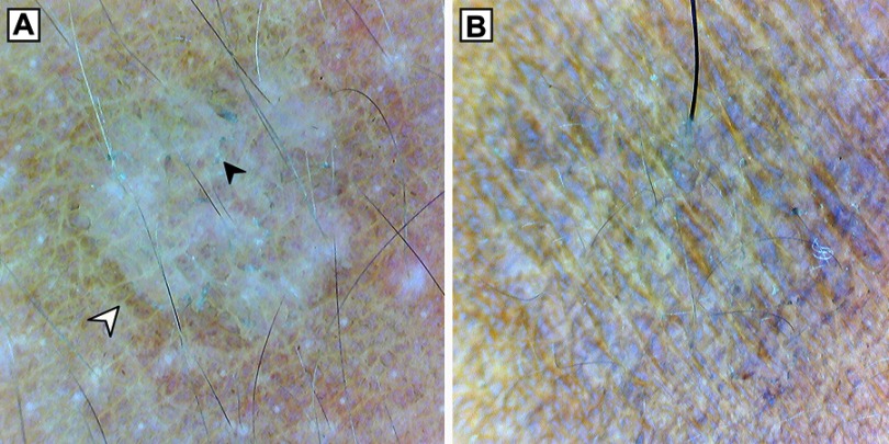
Figure 2.
(A) Dermoscopy (original magnification 10×) of hypopigmented lesion showing nonuniform pigmentation, inconspicuous ridges and furrows, perilesional hyperpigmentation (white arrowhead) and patchy scaling (black arrowhead). (B) Dermoscopy (original magnification 10×) of hyperpigmented lesion showing non-uniform pigmentation.

