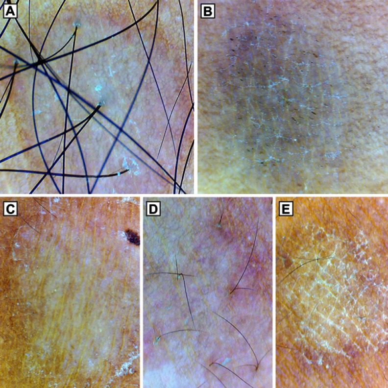
Figure 3.
Dermoscopy (original magnification 10×) showing different patterns of scaling in pityriasis versicolor. (A) Patchy scaling in hypopigmented lesion. (B) Diffuse scaling in hyperpigmented lesion with the simultaneous presence of scaling in the furrows. (C) Peripheral scaling. (D) Perifollicular scaling. (E) Scaling in the furrows. Note the nonuniform pigmentation in all the lesions, perilesional hyperpigmentation in (A) and inconspicuous ridges and furrows in (A), (D).

