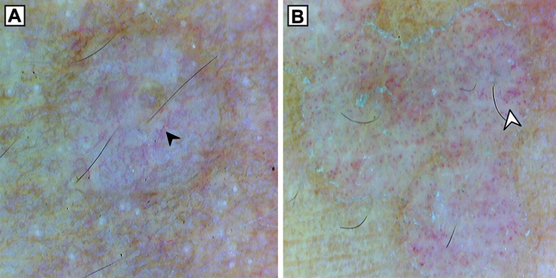
Figure 4.
Dermoscopy (original magnification 10×) showing different vascular patterns in pityriasis versicolor. (A) Linear branching vessels (black arrowhead). (B) Dotted vessels (white arrowhead). Note perilesional hyperpigmentation in (A), peripheral scaling in (B) and inconspicuous ridges and furrows in both (A) and (B).

