
Fig. 4.
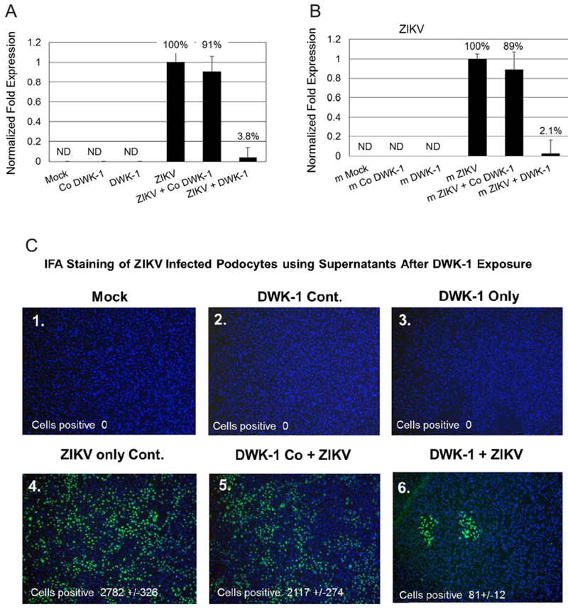
DWK-1 suppression of ZIKV progeny released from infected podocytes. (4A) qRT-PCR analysis of ZIKV replication in podocytes cultured under different conditions: 1. mock, 2. control morpholino, 3. DWK-1, 4. ZIKV, 5. control morpholino pretreatment and ZIKV, 6. DWK-1 pretreatment and ZIKV. Relative expression of intracellular ZIKV RNA was normalized to GAPDH mRNA. Values represent mean ± SD of 3 independent samples. *P < 0.001. ND, not detected. (4B) qRT-PCR analysis of ZIKV progeny released from podocytes cultured under different conditions: 1. mock, 2. control morpholino, 3. DWK-1, 4. ZIKV, 5. control morpholino pretreatment and ZIKV, 6. DWK-1 pretreatment and ZIKV. Relative expression of intracellular ZIKV RNA was normalized to GAPDH mRNA. Values represent mean ± SD of 3 independent samples. *P < 0.001. ND, not detected. (4C). ZIKV progeny released from infected podocytes by fluorescent focus infectivity assay using the 4 G-2 antibody specific to the E protein of ZIKV. (1) mock, (2) control morpholino, (3) DWK-1, (4) ZIKV, (5) control morpholino pretreatment and ZIKV, (6) DWK-1 pretreatment and ZIKV. All cells were stained with the 4 G-2 antibody conjugated FITC and counterstained with DAPI to stain the nuclei. Cells were counted by fluorescent microscopy. Fluorescent images were taken on a Nikon TE2000S microscope mounted with a CCD camera at 200 × magnification. Nuclei (blue) were stained with DAPI.