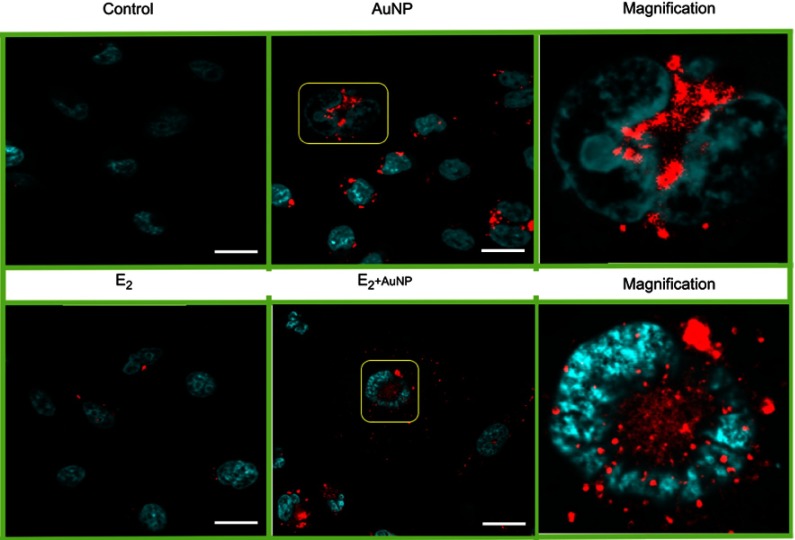
Figure 4.
Representative CLSM images of intracellular AuNPs after 12 h of incubation in MCF-7 cells. AuNP uptake is enhanced by E2 and particles are taken closer to the cell nucleus. The fluorescence signal of AuNP (red) was observed around the cell nuclei that were stained with DAPI (cyan). Digitally zoomed images (10× magnification) are shown in right column to better illustrate the localization of AuNP. Scale bars are 20 μm.
Abbreviations: AuNP, gold nanoparticle; CLSM, confocal laser scanning microscopy; DAPI, 4ʹ,6-diamidino-2-phenylindole.

