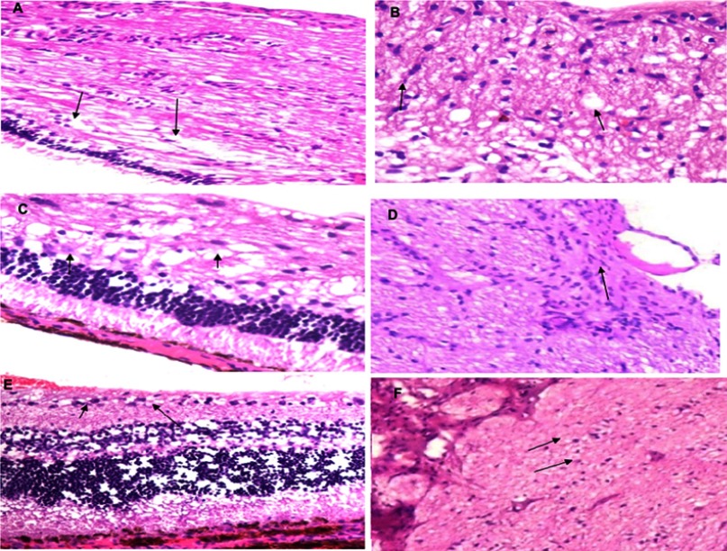
Figure 4.
Histopathological examination; findings were appointed with the arrows shown in each image. (A) Glaucoma retina (retina showing loss of internal nuclear layer and ganglion cells with numerous numbers of vacuoles in the choroid). (B) Glaucomatous optic nerve (optic nerve showing thickening, demyelination, and accumulation of fat vacuoles). (C) Glaucoma retina treated with acetazolamide (ACZ) oral tablet (retina showing some loss of inner plexiform layer and ganglion cells with little vacuoles in choroid). (D) Glaucomatous optic nerve treated with ACZ oral tablet (optic nerve showing Wallerian degeneration and minimal loss of myelin sheath). (E) Glaucoma retina treated with ACZ nanovesicles loaded in Chitosan nanogel (retina showing mild swelling of ganglion cells). (F) Glaucomatous optic nerve treated with ACZ nanovesicles loaded in Chitosan nanogel (optic nerve showing mild edema and thickening with remyelination of nerve fibers). Magnification power was 100×.

