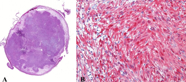
Figure 2.
Case 1. Histological feature exhibits A) low power transverse section through the mass showed a neurogenic, spindle cell proliferation with biphasic pattern of growth and prominent nuclear palisading. (hematoxylin and eosin stain, magnification 10×); B) The spindled cells arranged haphazardly within the loosely textured matrix. Classic pattern of Antoni B areas (Hematoxylin–eosin original magnification × 400)

