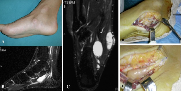
Figure 3.

Case 4. A) Clinical photograph of the foot in lateral, non–weight-bearing view shows the presence of swelling on the medial aspect of the right foot. B) Sagittal T2-weighted MRI scan with fat suppression revealing an hyperintense homogeneous fusiform mass on medial aspect of the foot along the course of the medial plantar nerve. C) Coronal T2-weighted MRI scan with fat suppression high enhancement of the lesion. D) Intraoperative localization of the mass in the medial part of the foot. E) It is clearly shown the connection with the medial plantar nerve
