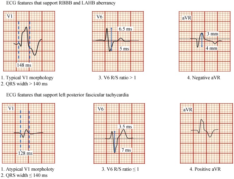
Figure 11. Schematic electrocardiogram representations.
Differentiating the QRS morphology of LPFVT from RBBB and LAHB aberrancy, when 3 or 4 criteria are positive the diagnosis of left posterior fascicular VT is likely. LAHB: left anterior hemiblock; LPFVT: left posterior fascicular ventricular tachycardia; RBBB: right bundle branch block; VT: ventricular tachycardia.

