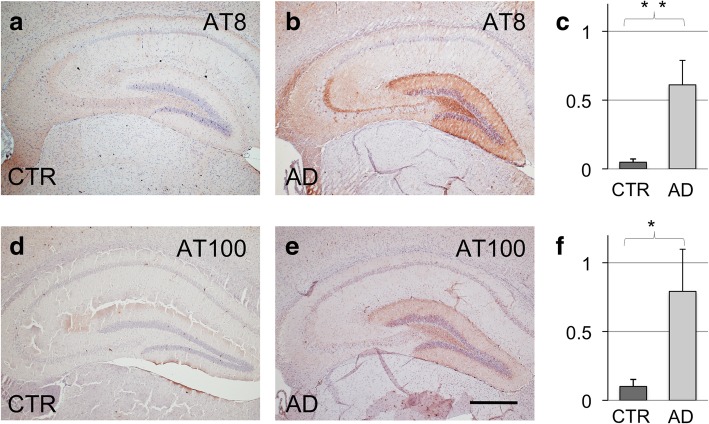
Fig. 1.
Immunohistochemistry for AT8 and AT100 tau phosphorylation markers of 7 months old P301S transgenic mice, sacrificed 4 months after unilateral hippocampal inoculation with CSF derived from control patients (CTR) or AD patients (AD) (a, d; b, e). AT8 positive hippocampal neurons (c, p = 0,001) ipsilateral to the inoculation site; AT100 positive hippocampal neurons, ipsilateral (f, p = 0,0013). Numbers indicate neurons per area. Scale bar in e equals 500 μm, and applies to a, b, d, and e

