Figure 3.
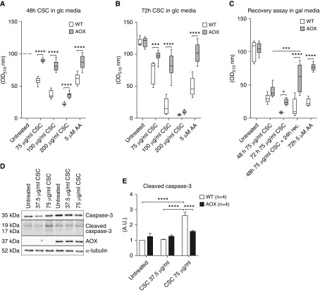
Analysis of CS condensate (CSC) toxicity in cultured immortalized mouse embryonic fibroblasts (iMEFs). (A–C) WT and AOX-expressing iMEFs cultured in glucose (glc, 10 mM) or galactose (gal, 10 mM) media and treated with CSC as indicated. Numbers of viable cells were estimated using the sulforhodamine B assay (absorbance at OD510) and normalized against the value at 48 hours for untreated cells on the given medium (shown in %). In C, cells recovered for 24 hours in gal (10 mM) without CSC as indicated after CSC (75 μg/ml) exposure. Horizontal bands inside the boxes represent the median (second quartile), the bottom and top of the box are the first and third quartiles, respectively, and the ends of the whiskers represent the minimum and maximum values of the data set. (D) Western blot of caspase-3, cleaved caspase-3, and AOX, with α-tubulin as the loading control. For entire blots, including molecular-weight markers for cropped Western blot bands, please refer to Figure E5. (E) Relative densitometric analysis (n ≥ 3) on proteins extracted from WT and AOX iMEFs exposed to CSC as indicated in gal media. Bar graph represents mean ± SEM; *P < 0.05, ***P < 0.0005, and ****P < 0.0001 by two-way ANOVA. AA = antimycin A; OD = optical density.

