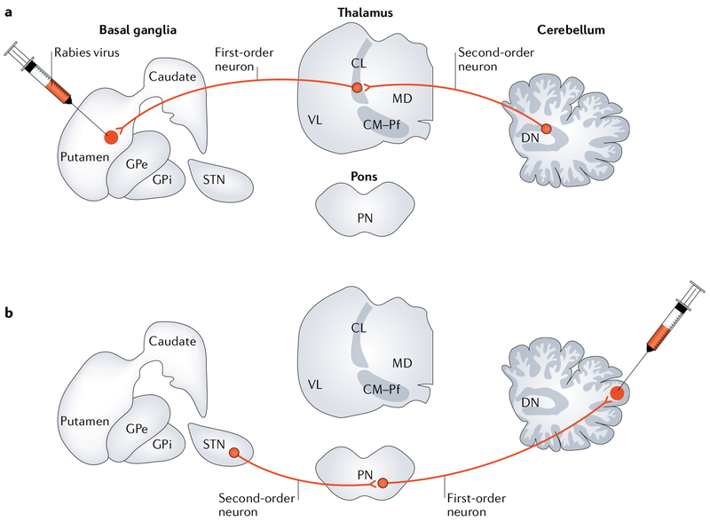
Fig. 2 |. Anatomical connections.
a | A study using retrograde transneuronai transport of rabies virus in monkeys revealed a disynaptic pathway from the dentate nucleus (DN) to the putamen11. Rabies virus was injected into the putamen and underwent retrograde transport to first-order neurons that project to the striatum (for example, neurons in the intralaminar thalamic nuclei) and then retrograde transneuronal transport to second-order neurons that innervate the first-order neurons. The second-order neurons in the cerebellum were located primarily in the DN. b | A study using retrograde transneuronal transport of rabies virus in monkeys revealed a disynaptic pathway from the subthalamic nucleus (STN) to the cerebellar cortex12. Rabies virus was injected into the lateral cerebellar cortex and underwent retrograde transport to first-order neurons that project to the cerebellar cortex (for example, neurons in the pontine nuclei (PN)) and then retrograde transneuronal transport to second-order neurons that innervate the first-order neurons. The study revealed second-order neurons labelled in the basal ganglia, primarily in the STN. CL, central lateral thalamic nucleus; CM, central medial thalamic nucleus; GPe, external segment of the globus pallidus; GPi, internal segment of the globus pallidus; MD, medial dorsal thalamic nucleus; Pf, parafascicular thalamic nucleus; VL, ventral lateral thalamic nucleus.

