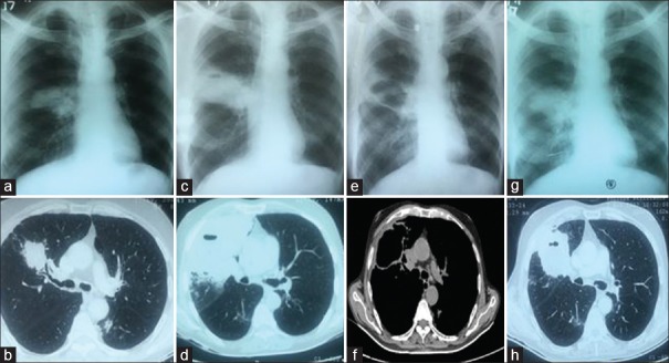
Figure 1.
(a) Chest radiograph at presentation to us showing increase in the size of the right hilar region compared to previous radiograph shown in . (b) Chest computed tomography scan lung window, transverse cut at the level of carina, corresponding to the chest radiograph in a showing mass with tumor bronchus sign. (c) Chest radiograph after 1 month of a showing further increase in the size of the right hilar lesion. (d) Chest computed tomography scan lung window transverse cut at the level of carina, at the time of first computed tomography-guided biopsy corresponding with radiograph of c showing mass in the right upper lobe extending up to the pleura. (e) Chest radiograph after 1 month of c showing a cystic opacity at the site where initially solid lesion was seen. (f) Corresponding computed tomography scan to the chest radiograph shown in e, mediastinal window at the level of carina showing cystic opacity and regression of lesion. (g) Chest radiograph almost 3 months after nephrectomy showing re appearance of the lesion at the same site. (h) Corresponding computed tomography scan to the chest radiograph in g, lung window at the level of carina showing solid mass in right upper lobe

