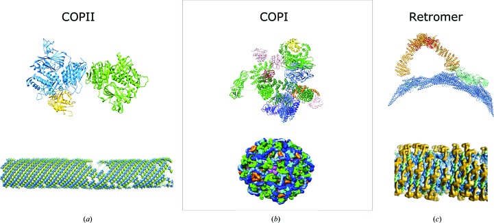
Figure 5.
Structural insights gained from the study of coat proteins. The top panels represent fitted structural models of asymmetric units. The bottom panels show the global coat arrangement. (a) Inner COPII coat (Hutchings et al., 2018 ▸; PDB entry 6gni). Colour scheme: Sec23, blue; Sec24, green; Sar1, yellow. (b) COPI (Dodonova et al., 2015 ▸, 2017 ▸; PDB entry 5nzr). Colour scheme: Arf1, pink; γ-COP, light green; β-COP, dark green; ζ-COP, yellow; δ-COP, orange; β′-COP, light blue; α-COP, dark blue. (c) Retromer (Kovtun et al., 2018 ▸; PDB entry 6h7w). Colour scheme: Vps5, blue; Vps29, red; Vps35, yellow; Vps26, green. The images are reproduced with permission from AAAS and Springer Nature.

