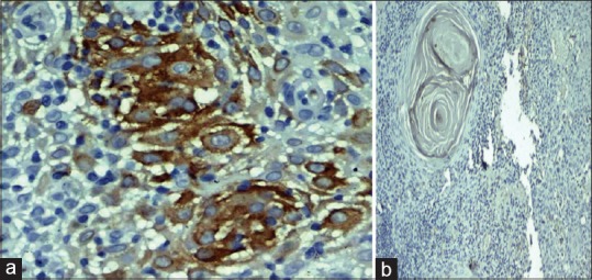
Figure 2.

(a) Photomicrograph showing intense membrane and cytoplasmic staining of MUC4 in the keratin pearl of well-differentiated squamous cell carcinoma (IHC, ×40). (b) Absence of MUC4 expression in the keratin pearl of well-differentiated squamous cell carcinoma (IHC, ×40)
