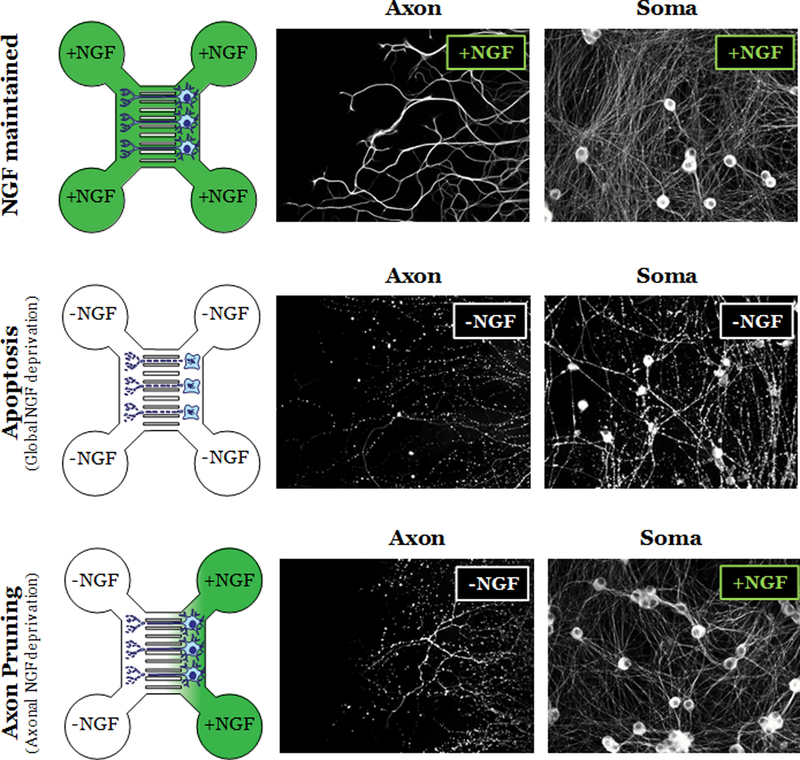
Fig. 1. Microfluidic chamber model for studying the pathways of apoptosis and axon pruning.
(Left) Illustration of microfluidic chambers in NGF maintained (top), Apoptosis (global NGF deprivation; middle), and Axon pruning (axonal NGF deprivation; bottom) conditions. (Right) Fluorescent images of neurons labeled with α-tubulin shows healthy and degenerated soma and axons in the indicated conditions.

