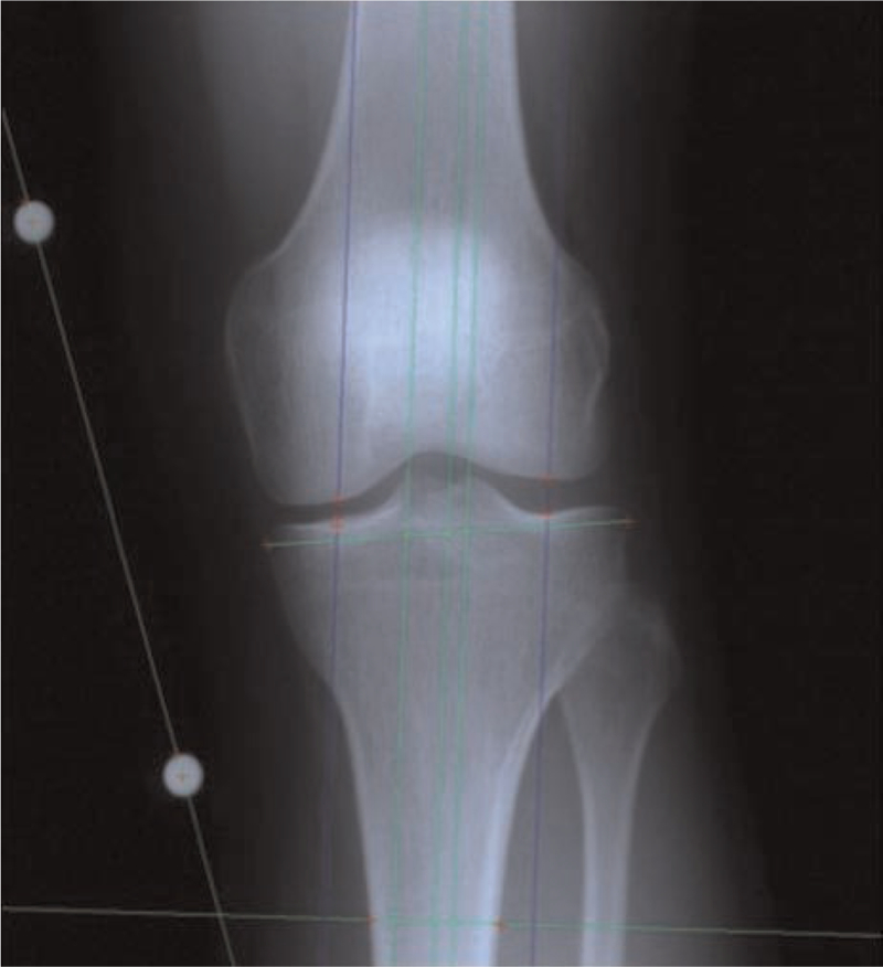
Figure 2.

Scanned radiograph analyzed with a semiautomated technique. Calibration beads adhering to the medial aspect of the knee were used to adjust for magnification and to normalize measures between radiographs. A reference line was drawn through the midpoints of lines drawn across the tibial plateau and tibial shaft (at 1 times the distance of the tibial plateau line length down the tibia from the tibial plateau line). All additional lines were drawn in parallel to this line. The most prominent peaks of the tibial spines were identified, and medial and lateral compartment “mid-point” lines were drawn halfway between the tibial border and its respective tibial spine. Joint space width measurements were made in each compartment at the location of these midpoint lines.
