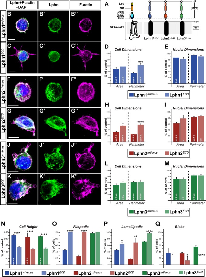
Fig. 2.
Uncoupling Lphns from the intracellular machinery distinguishes between NTF- and CTF-dependent actin remodeling functions. (A) Schematic representation of LphnECD proteins containing both HA and myc epitopes followed by the transmembrane domain of platelet-derived growth factor receptor (TM*). Represented domains are: lectin (Lec), olfactomedin (Olf), hormone binding (HRM), GPCR auto-proteolysis inducing (GAIN), GPCR proteolysis site (GPS), seven transmembrane domains and interconnecting loops (GPCR-like), N-terminal fragment (NTF). (B,C,F,G,J,K) HEK293T cells expressing indicated proteins were visualized by confocal microscopy after staining for nuclei (in blue), for F-actin (in magenta) and HA epitope or mVenus fluorescence (in green). (D,E,H,I,L,M,N) Cell and nuclei dimensions as well as cell height values represented as a percentage of mVenus-expressing cells values. (O,P,Q) Percentage of cells harboring filopodia, lamellipodia or blebs. Scale bars: 10 µm. Data, represented as mean values, were obtained from at least three separate experiments (Ctrl n=23, Lphn1mVenus n=27, Lphn1ECD n=29, Lphn2mVenus n=27, Lphn2ECD n=34, Lphn3mVenus n=30, Lphn3ECD n=30). Error bars indicate s.e.m. Gray asterisks indicate significance with control values from cells expressing mVenus. ****P≤.0001, ***P≤0.001, **P≤0.01, *P≤0.05.

