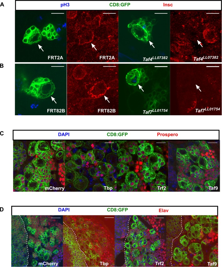
Fig. 4.
NSC-TAF or TRF2-depleted NSCs exhibit defective cell polarity but do not express differentiation markers. (A) Taf4LL07382 NSCs or (B) Taf7LL07382 NSCs fail to express normal levels of Insc. Mitotic cells are labeled with phospho Histone H3 (pH3), CD8:GFP (a membrane-tethered GFP) labels the clones and Insc is shown in red. (C) Knockdown of TBP, TRF2 or TAF9 does not result in nuclear accumulation of Prospero. (D) Knockdown of TBP, TRF2 or TAF9 does not result in nuclear accumulation of Elav. The white dotted line demarcates the optic lobe (left)/central brain (right) boundary. In both C and D, transgenes were expressed using a UAS-Dcr2; inscGAL4, UAS-CD8:GFP, tubGAL80ts driver and transgenes induced for 72 h. Scale bars: 10 µm (A–C); 20 µm (D).

