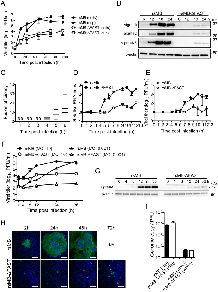
Fig 2. FAST protein enhances viral replication at an early phase of infection.
(A) Vero cells were infected with rsMB or rsMB-ΔFAST at a multiplicity-of-infection (MOI) of 0.01 plaque-forming units (PFU)/cell. Infectious virus titers in the supernatant and cell lysate were examined. Data are expressed as means ± SD (n = 3). (B) Time course of viral protein expression. Vero cells were infected with rsMB or rsMB-ΔFAST at a MOI of 0.01 PFU/cell. Whole-cell extracts were subjected to western blotting. Viral proteins were detected with antibodies to sigmaC, p17, and sigmaA. (C–E) Vero cells were infected with rsMB or rsMB-ΔFAST at a MOI of 0.01 PFU/cell and incubated for various intervals. (C) To determine the time course of syncytium formation, cells were fixed and viral sigmaC antigens were visualized by immunostaining. Numbers of cells involved per syncytium were plotted. Data are presented as a box plot (n = 11–30). (D, E) To determine the kinetics of viral replication, cells were disrupted at indicated times post infection by freeze-thawing. Copy numbers of the genomic RNA L1 gene (D) and infectious virus titers (E) were investigated by TaqMan quantitative PCR and plaque-formation assay, respectively. Arrows indicate the time points of primary increase of levels of viral genomes and infectious virus particles. NA, not available; ND, not detected. Data are expressed as means ± SD (n = 3). (F, G) Vero cells were infected with rsMB or rsMB-ΔFAST at a MOI of 0.001 or 10 PFU/cell. (F) Virus infectious titers were determined by the plaque assay. (G) Viral antigens in whole-cell extracts of Vero cells infected with rsMB or rsMB-ΔFAST at a MOI of 10 PFU/cell were detected with an anti-sigmaA antibody. An anti-β-actin antibody was used as a loading control. (H) Time course of virus spread in monolayers of Vero cells. Vero cells were infected with rsMB or rsMB-ΔFAST at a MOI of 0.01 PFU/cell. After virus adsorption at 37°C for 1 h, cells were overlaid with 0.8% agarose gel and incubated. At the indicated time points post infection, cells were fixed and viral antigens were detected by immunostaining with murine anti-PRV-MB serum followed by anti-mouse IgG-CF488. NA, not available. Scale bars are 200 μm. (I) Ratio between the viral genome copy number and the infectious virus titer in whole-cell lysates and purified virions. The genome copy number of positive-stranded L1 gene segments and the number of plaque-forming units of purified rsMB and rsMB-ΔFAST were calculated. Data are expressed as means ± SD (n = 3) and were statistically analyzed using the t-test.

