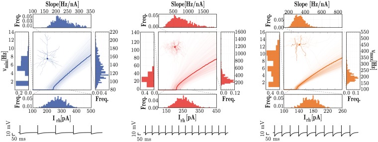
Fig 1. Response properties of the three different neuronal types, E (left), I1 (middle) and I2 (right).
For each neuronal class, the central panels depict single neuron fI curves and the marginal panels give the corresponding distributions of the neuron’s rheobase currents (Irh[pA], bottom), minimum firing rates (νmin[Hz], left); maximum firing rates (νmax[Hz], right) and the slope of the fI curve (Slope[Hz/nA], top). The data was obtained from 1000 neurons of each class. The membrane potential traces depicted in the bottom correspond to the response of the homogeneous neurons (bold traces in the fI curves) to a stimulus step of amplitude Irh + 10 pA, for a duration of 1 second.

