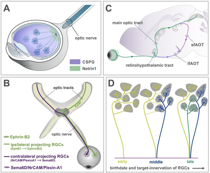
Figure 1: RGC axon pathfinding and target selection.
(A) Retinal ganglion cells extend axons away from the periphery due to repulsive influences from chondroitin sulfate proteoglycans (CSPG, purple) and grow towards the optic disk. Netrin 1 expressed by glial cells at the optic disc (green) provides local attractive cues and enables axons to exit the eye, into the optic nerve.
(B) Schematic of the optic nerve reaching the optic chiasm, a midline choice point for RGC axons. EphrinB2 expressed at the midline repels EphB1 expressing RGC axons to form ipsilateral projections (green), while Sema6D/NrCAM/PlexinA1 complex directs other RGCs to cross at the chiasm and form the contralateral projections (magenta).
(C) Schematic showing the anatomical position of the eye, optic nerve, and optic tracts projecting to targets in the brain. After crossing the chiasm, the contralateral projection ascends into the brain to form the main optic tract (green). A smaller bundle projects into the SCN at the base of the hypothalamus, forming the retinohypothalamic tract. Two bundles deviate from the tract however – the inferior fasciculus of the accessory optic tract (ifAOT, purple), extends at the base of the brain to project to the MTN. While another bundle continues from the main optic tract and dives down to form the superior fasciculus of the AOT (sfAOT, pink).
(D) Birth order of RGCs determines their target-selection and exploration. Early-born RGCs extend axon branches into many targets (yellow lines). Axons born shortly after (blue) extend to a few different targets while the early born axons retract some connections (yellow dotted lines). The later-born axons project directly to their targets without much exploration (green).

