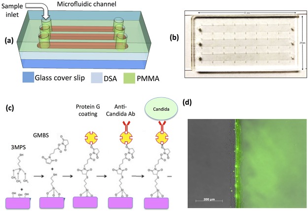
Figure 2.
(a) Schematic of the microfluidic chip consisting of PMMA, DSA, and glass cover. (b) Actual image of the assembled microchip containing three microfluidic channels, inlets, and outlets. (c) Protein G-based surface chemistry was used to immobilize anti-Candida antibodies on the surface. (d) Fluorescent image showing clear green signal (right side of microchannel) that indicates successful immobilization of fluorescein isothiocyanate (FITC)-conjugated protein.

