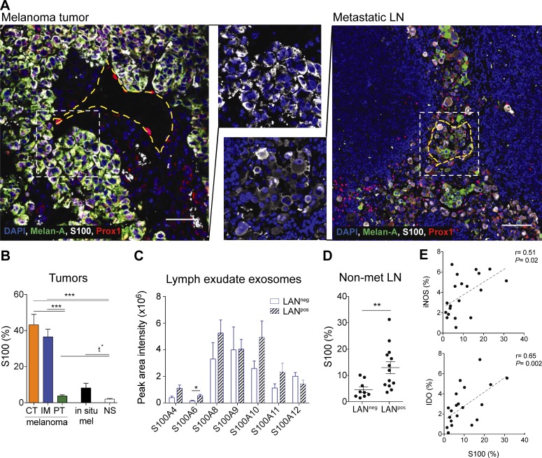
Figure 6.
Premetastatic LNs express S100 proteins that are found in lymphatic exudate EVs of melanoma patients. (A) Primary melanoma (left; bar, 50 µm) and metastatic LN (right; bar, 200 µm) analyzed by immunofluorescence staining. Yellow dotted lines indicate the perimeter of lymphatic vessels (identified by nuclear Prox-1 staining) while the white dotted lines indicate the zoomed areas shown in the center. (B) Quantification of S100 (% of positive pixels) in different regions of the melanoma (CT, center of the tumor; IM, invasive margin; PT, peritumoral region; n = 14) compared with in situ melanoma (mel; also called stage 0 melanoma, which is confined to the epidermis; n = 14) and normal skin (NS, n = 12). (C) Expression of S100 proteins in EVs isolated from lymphatic exudate of melanoma patients as analyzed by mass spectrometry. (D) S100 expression in whole sections of tumor-free (nonmetastatic [non-met]) LNs of LANneg (n = 10) and LANpos (n = 12) patients. Data shown as mean ± SEM. (E) Correlation of S100 expression with iNOS and IDO expression in the tumor-free LNs of both LANpos and LANneg patients (n = 23).

