Figure 8.
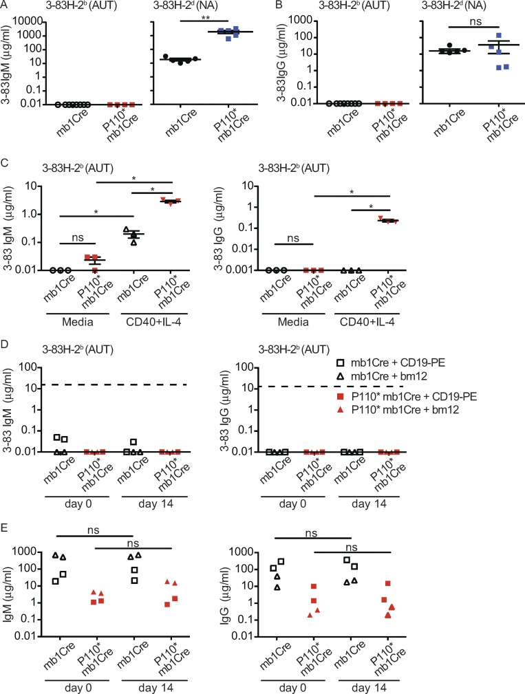
Activation of PI3K causes production of high-avidity autoantibodies in response to T cell help in vitro, but not in vivo. (A and B) Concentration (μg/ml) of 3–83IgM (A) and 3–83IgG (B) in sera of mb1Cre and P110*-mb1Cre mice on either the autoreactive (AUT) 3–83Igi,H-2b (n = 8 and 4) or the nonautoreactive (NA) 3–83Igi,H-2d (n = 5) backgrounds analyzed in two experiments. (C) Concentration (μg/ml) of 3–83IgM and 3–83IgG in the supernatant of splenic B cells from autoreactive mb1Cre and P110*-mb1Cre mice cultured for 8 d in either media alone or in the presence of anti-CD40 antibodies and IL-4. n = 3 per group from two independent experiments. (D and E) Concentration (μg/ml) of 3–83IgM and 3–83IgG (D) and total IgM and IgG (E) in the sera of autoreactive mb1Cre or P110*-mb1Cre mice before (day 0) and on day 14 after the injection of either anti-CD19-PE antibodies or the transfer of bm12 (B220−) spleen cells. n = 2 for each experimental condition, representative of two independent experiments. The dashed lines in D indicate the reference levels of 3–83IgM and 3–83IgG in 3–83Igi,H-2d mice. Data in A–C show the mean ± SEM. Data were analyzed using a one-tailed Mann–Whitney U test. *, P ≤ 0.05; **, P ≤ 0.01; ns, not significant.

