. 2019 Apr 29;8:e44328. doi: 10.7554/eLife.44328

Figure 6. Atoh1 overexpression in utricle supporting cells can remodel chromatin of hair cell gene loci.

(A) Aggregation plot showing enrichment of ATAC-seq signals from each condition for unique utricle peaks that are assigned to the 945 hair cell genes that can be induced in supporting cells (left) or the 967 hair cell genes that cannot be induced in supporting cells (right). No significant differences are seen in the relative accessibility of these two groups of hair cell genes. (B) Example of an ATAC-seq profile for a hair cell-specific gene, Rasd2 that can be altered by Atoh1 transduction. RNA-seq analysis shows that Rasd2 is up-regulated in supporting cells by transduction with Atoh1, but not with the TdTomato control virus. A unique ATAC-seq peak present in hair cells but not supporting cells (yellow highlighting). However, the peak re-appears in supporting cells transduced with Atoh1, but not with the TdTomato control virus. The DNA sequence corresponding to this peak shows binding sites for Atoh1 along with other transcription factors. (C) Example of ATAC-seq profiles for a hair cell-specific gene, Kcnj13. RNA-seq analysis shows that Kcnj13is not up-regulated in supporting cells by transduction with Atoh1 or with the TdTomato control virus. Nevertheless, a unique ATAC-seq peak present in hair cells is re-opened in supporting cells transduced with Atoh1.

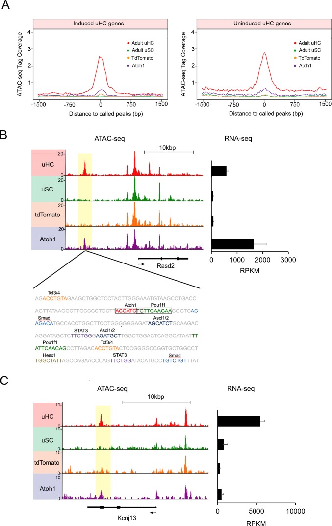
Figure 6—source data 1. Known motif analysis of transcription factor binding sites that are enriched in the accessible chromatin of utricle hair cells, utricle supporting cells, neonatal cochlear hair cells, Ad-tdTomato or Ad-Atoh1 infected supporting cells.
DOI: 10.7554/eLife.44328.024

Figure 6.

Figure 6—figure supplement 1. Analysis of hair cell gene up-regulation in supporting cells.

Figure 6—figure supplement 1.

(A) Venn diagram showing identification of hair cell genes that are up-regulated in supporting cells by Atoh1 transduction (945 genes) compared to hair cell genes that failed to be activated by Atoh1 (967 genes). (B) Analysis of ATAC-seq data from 1912 utricle hair cell gene loci showing the proportion of loci containing accessible peaks in genes that were induced by Atoh1 versus those that were not induced by Atoh1. TSS: transcription start site.
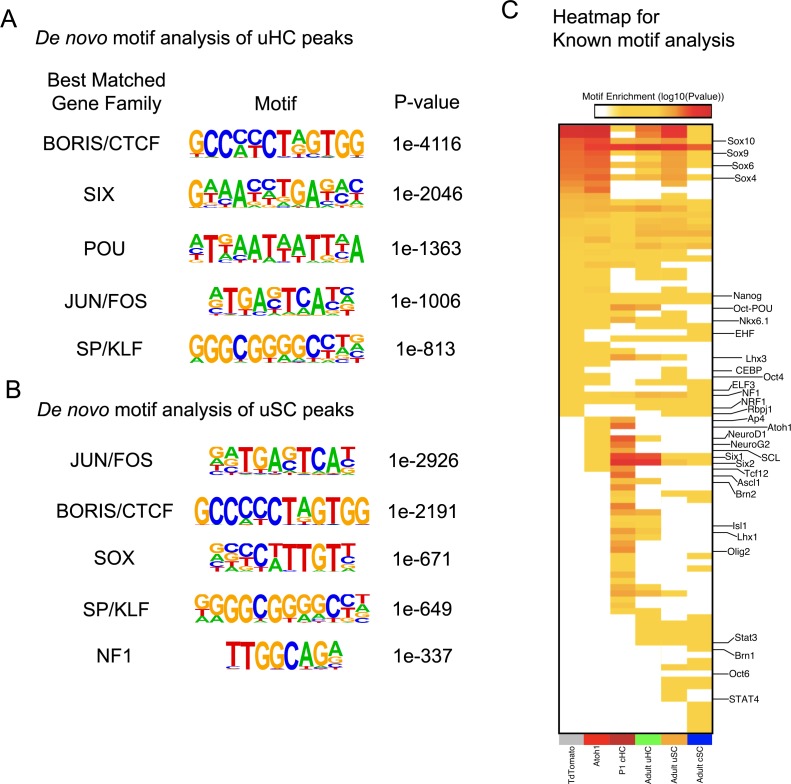
Figure 6—figure supplement 2. Motif enrichment analysis showing of utricle hair cells and supporting cells, and changes induced by Atoh1 transduction.

Figure 6—figure supplement 2.

(A) De novo motif analysis of all called ATAC-seq peaks from purified utricle hair cells. The top five transcription factor binding motifs discovered in the peaks are shown. Note that Atoh1 is not expressed in mature hair cells, therefore Atoh1 binding motifs were not significantly present in the utricle hair cell peaks. (B) De novo motif analysis of all called ATAC-seq peaks from purified utricle supporting cells. The top five transcription factor binding motifs discovered in the peaks are shown. (C) Heat map showing the relative enrichment of transcription factor binding motifs in called ATAC-seq peaks in utricle hair cells, utricle supporting cells, neonatal (P1) cochlear hair cells, neonatal (P1) cochlear supporting cells, adult (P21) cochlear supporting cells and utricle supporting cells infected with either Ad-tdTomato or Ad-Atoh1. Atoh1 transduction causes an increase in accessible peaks enriched for binding sites in Atoh1 itself (Figure 6C), NeuroD1, Neurog2, SCL, Six1, Six2 and Tcf12.
Figure 6—figure supplement 3. Atoh1 overexpression induced chromatin accessibility is independent for up regulation of hair cell genes in supporting cells.

Figure 6—figure supplement 3.

(A) Quantification of percentage of utricle hair cells specific peaks became accessible in Ad-Atoh1 infected supporting cells for induced or uninduced uHC genes’ loci. (B) De novo motif analysis identified transcription factor binding sites in the utricle hair cell-specific accessible chromatin that became accessible and were expressed in Ad-Atoh1 infected supporting cells. (C) De novo motif analysis also identified transcription factor binding sites in the utricle hair cell specific accessible chromatin that became accessible in Ad-Atoh1 infected supporting cells, but whose associated genes were not expressed after Atoh1 transduction.
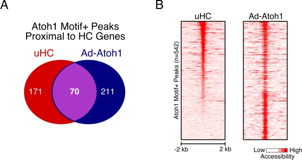
Figure 6—figure supplement 4. Atoh1 Overexpression Promotes Ectopic Peak Activation Proximal to Hair Cell Genes.

Figure 6—figure supplement 4.

(A) Venn diagram showing the overlap of Atoh1-motif containing peaks identified proximal to HC genes activated by Ad-Atoh1 for uHCs and Ad-Atoh1 cells. (B) Heatmap showing ATAC-seq signal across the peaks shown in (A).