. 2019 Feb 18;8:e40472. doi: 10.7554/eLife.40472

Figure 2. Mechanics of binding.

(A) A schematic of the model. A protein filament (red) may bend and bind to a cylindrical membrane (green) at an angle with respect to the long axis, θ, and the equilibrium conformation may involve deformations of both protein and membrane. (B) Plot of the estimated free energy change due to filament binding against the binding angle, θ, where the cell radius Rcell=0.5μm, the pressure difference across the membrane p=20atm, the estimated turgor pressure of B. subtilis (Whatmore and Reed, 1990), and the other model parameters are given in Supplementary file 1. The estimate is similar for p=0.2atm, but exhibits a more shallow potential well for Rcell=1.5μm (red curve). (C) An approximate phase diagram of protein-membrane binding. The dashed lines delineate regimes, as explained further in Appendix 1.

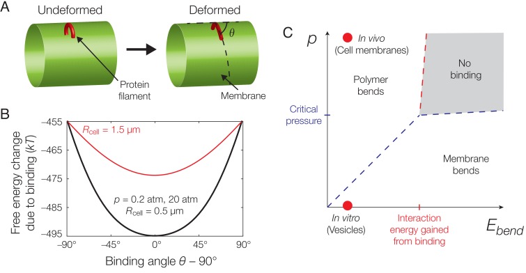
Figure 2.

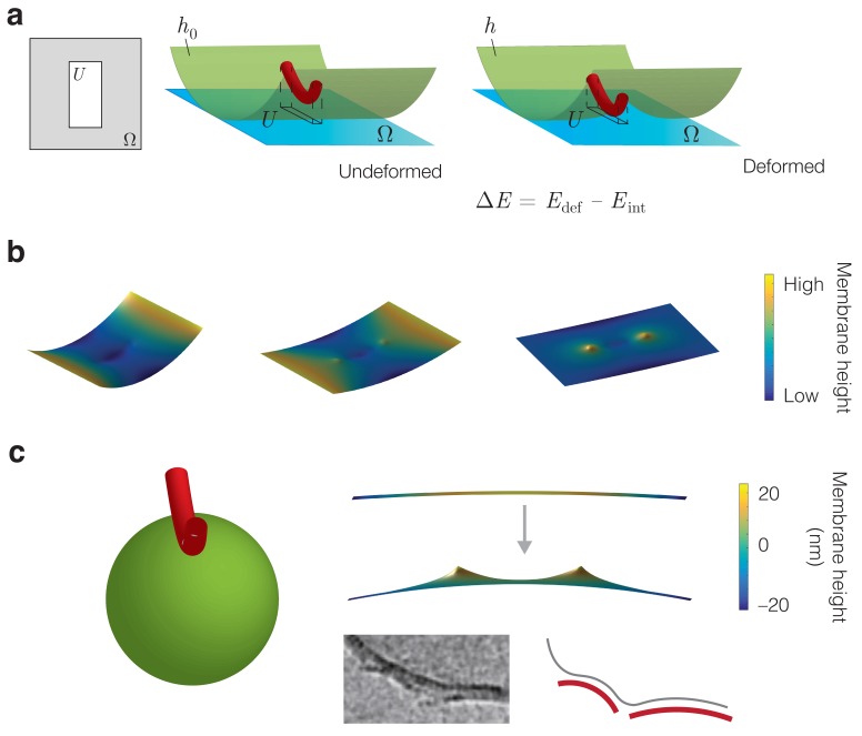
Figure 2—figure supplement 1. Energetic modeling of MreB binding and deformation of vesicles.

Figure 2—figure supplement 1.

(a) A protein filament is modeled as a curved elastic rod that attaches to the inner membrane in an energetically favorable manner. U denotes the interaction region. In the Monge gauge, a height function h(x,y) parameterizes the membrane shape (h0 in the undeformed state) and the domain of h (h0) is denoted Ω. The interaction region restricts the value of h over the region U. (b) Example solutions for the membrane shape for p=0 and Rcell=0.5 μm, 1.5 μm, and (left to right) on a finite domain, for the parameter values summarized in Supplementary file 1. (c) (Left) The binding of an MreB filament to a spherical vesicle induces vesicle curvature, a schematic of which is shown. (Right top) A numerical solution for the membrane shape over a finite domain where the undeformed configuration is a sphere with a principal curvature opposite that of an MreB filament. Here p=0, the sphere radius Rsphere=1μm, and the remaining parameter values are summarized in Supplementary file 1. (Right bottom) MreB filaments binding to vesicles in vitro, with a schematic shown. The image is reproduced here from Salje et al. (2011) under a CC BY 3.0 license (https://creativecommons.org/licenses/by/3.0/).