Figure 1.

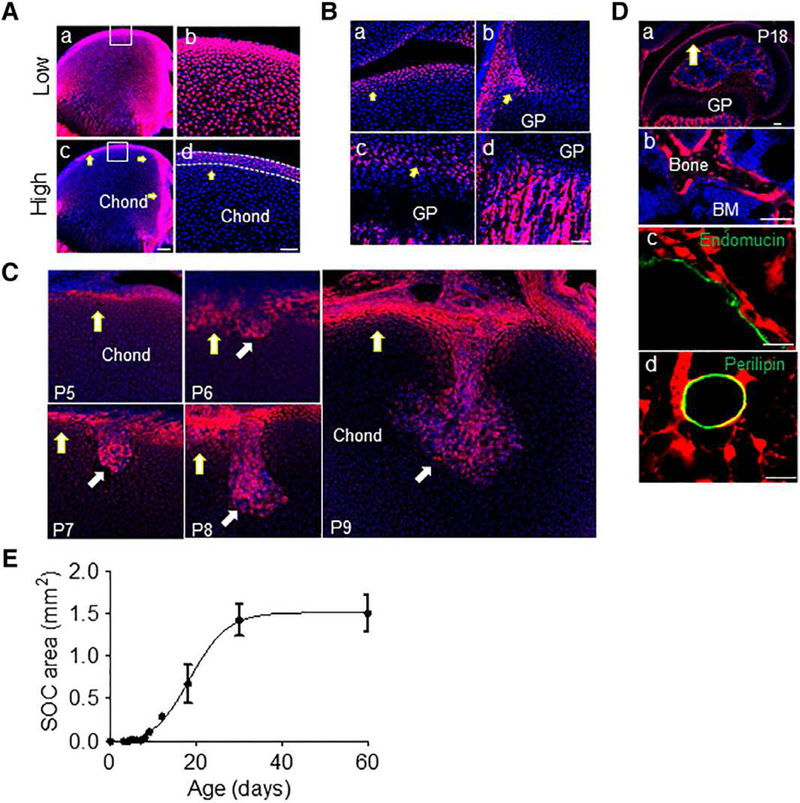
The highly fluorescent periarticular region in Col2/Tomato mice contains multipotent cells that forms the invading canal and reconstitutes subchondral bone. (A): A femur from P4 Col2/Tomato mouse was imaged by confocal microscopy and displayed at low (Aa, Ab) and high (Ac, Ad) intensity threshold. Yellow arrows point to TomatoH cells. The periarticular region was defined as the 4–6 layers of TomatoH cells along the epiphyseal surface (white dotted lines). Blue: DAPI. Bar = 200 μm (Aa, Ac), 50 μm (Ab, Ad). (B): High magnification images of the periarticular region (Ba), groove of Ranvier (Bb), growth plate (arrows point to the resting zone; Bc), and primary spongiosa (Bd) of the same femur showing labeling with high Tomato fluorescence. Bar = 50 μm. (C): Col2/Tomato mice were sacrificed postnatally at days indicated and the forming cartilage canal imaged by confocal microscopy. Bar = 100 μm. (D): By P18 (Da), TomatoH cells were found to give rise to osteoblasts and osteocytes (Db), pericytes along Endomucin+ blood vessels (Dc), and adipocytes (Dd). (Db), (Dc), and (Dd) are high magnification images from (Da). Bar = 200 μm (Da, Db), 25 μm (Dc, Dd). (E): Monitoring of secondary ossification center expansion revealed rapid growth during the invasion and expansion phases, with a maximum epiphyseal marrow area being reached by P30. Values represent averages ± SEM (n = 3–5/age).
