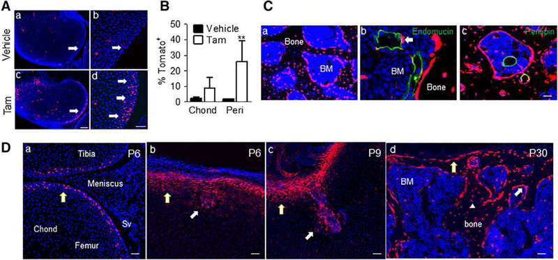
Figure 2.

Lineage tracing specifically labels periarticular cells which give rise to the invading canal and reconstitutes subchondral bone. (A): Tomato+ cells were predominantly found at the periarticular region (pointed by arrows) in Col2ER/Tomato mice at P5 after tamoxifen injection at P4. (Ab) and (Ad) are magnified images from (Aa) and (Ac), respectively. Bar = 200 μm (Aa and Ac), 50 μm (Ab and Ad). (B): Quantification of the number of Tomato+ cells in the chond and peri regions following tamoxifen injection; n = 3. Values represent average values ± SEM. **, p <.01 relative to vehicle control by two-way analysis of variance with Bonferroni’s post-test. (C): These mice were also found to have Tomato+ osteoblasts, osteocytes (Ca), perictyes (Cb), and adipocytes (Cc) at P60. Bar = 50 μm. (D): Gli1-CreER/ Tomato mice were injected with tamoxifen at P4 and sacrificed during canal initiation (P6), canal invasion (P9) and following secondary ossification center (SOC) formation (P30). Tomato signal was found to specifically label periarticular cells, the cartilage canal and mesenchymal lineage cells within the bone marrow following SOC formation, consistent with the TomatoH cells in the Col2/Tomato mice. Bar = 50 μm. Abbreviations: BM, bone marrow; Chond, Epiphyseal chondrocytes; Peri, periarticular; SV, synovium.
