Figure 3.

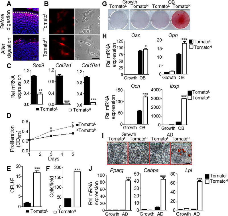
The periarticular region contains mesenchymal progenitors with increased osteogenic and adipogenic potential. (A): TomatoH and TomatoL cells were isolated by short-term or overnight enzymatic digestion, respectively. Bar = 50 μm. (B): Morphologically, TomatoL cells appear more rounded, characteristic of chondrocytes, whereas TomatoH cells appear more fibroblastic and elongated with increased fluorescence following adhesion. Bar = 10 μm. (C): Gene expression confirmed TomatoL cells expressed elevated levels of the chondro-genic markers Sox9, Col2, and Col10, relative to TomatoH cells. (D): TomatoH cells possessed significantly greater proliferation, as assessed by MTT assays. (E): TomatoH possess an increased colony forming ability. (F): TomatoH cells show increased migration rates, as assessed by trans-well assays. (G): TomatoH cells showed increased mineralization under osteogenic differentiation conditions. (H): TomatoH cells showed increased expression of the osteogenic markers Osx, Opn, Ocn, and Ibsp, relative to TomatoL cells, under osteogenic differentiation conditions. (I): Under adipogenic differentiation conditions, TomatoH cells show increased staining for lipid accumulation. Bar = 10 μm. (J): TomatoH cells expressed significantly elevated levels of the adipogenic markers Pparγ, Cebpa, and Lpl when cultured under adipogenic conditions. Graphs represent average values ±SEM. Data was analyzed by paired t test (C-F) or by repeated measures two-way analysis of variance with Bonferroni’s post-test (*, p <.05; **, p <.01; ***, p <.001; n = 3).
