Figure 5.

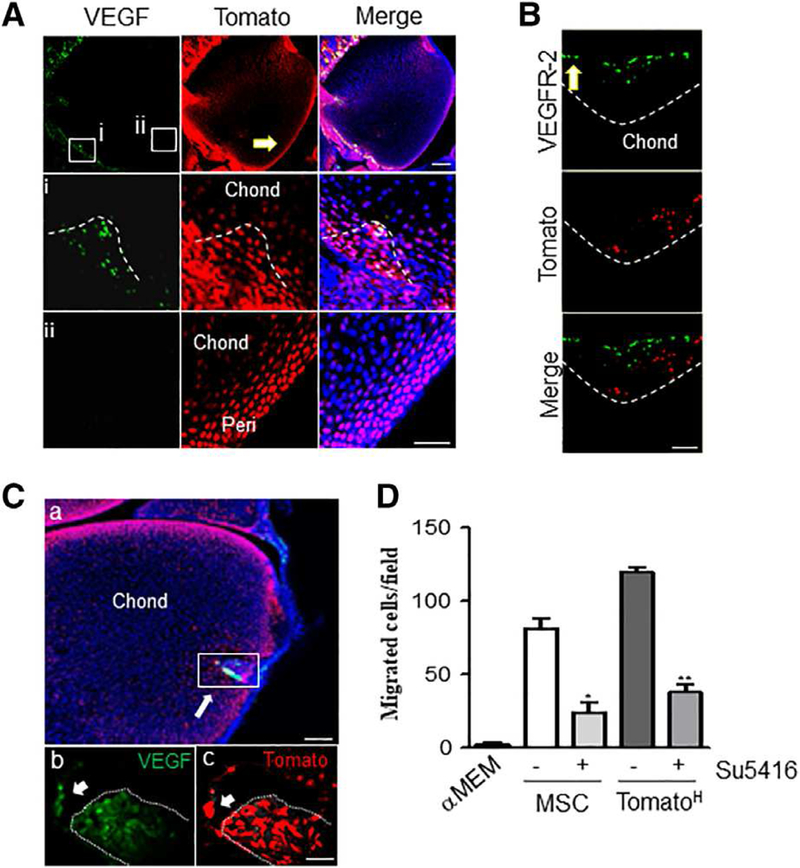
Secondary ossification center canal initiation and progression is led by perichondrial-derived Tomato cells independent of epiphyseal chondrocyte-derived vascular endothelial growth factor (VEGF) expression. (A): At P5, VEGF expression is confined to cells forming the canal and the neighboring periarticular region (Ai) while no signal was detected along the periarticular surface away from the canal (Aii). Dotted line depicts canal invasion site. Bar = 100 μm. (B): These leading cells are closely followed by a wave of VEGFR-2+ cells. Dotted line depicts canal front. Bar = 100 μm. (Ca): At P8, VEGF is expressed robustly by most cells of the canal, as well as a few chondrocytes in front of the advancing canal. Bar = 200 μm. (Cb, Cc): Magnified insert from (Ca). Arrow denotes TomatoH cells in front of canal positive for VEGF staining. Bar = 20 μm. (D): Trans-well migration of endothelial cells was conducted using conditioned media from mesenchymal stem cells orTomatoH cells and treatment with the VEGFR-2 inhibitor Su5416. TomatoH cells were found to induce endothelial cell migration in a VEGR-2-dependent manner. Graphs represent average values ± SEM. Data analyzed by repeat measures two-way analysis of variance with Bonferroni’s post-test (*, p <.05; **, p <.01; n = 3).
