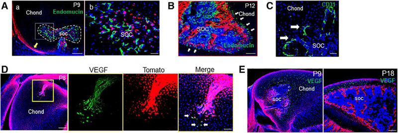
Figure 6.

The transition from canal invasion to secondary ossification center (SOC) expansion is associated with altered sources of vascular endothelial growth factor (VEGF) and a spatial reorganization of blood vessels. (A): By P9, some mature vessels were visualized and present along the front of the expanding SOC canal (dotted line). Yellow arrow denotes periarticular surface. Bar = 200 μm (Aa), 50 μm (Ab). (B): By P12, endothelial cells were found to be ahead of the expanding TomatoH bone compartment (arrows). Bar = 200 μm. (C): These vessels were associated with a high density of angiogenic sprouts (arrows) along the invading front. Bar = 50 μm. (D): This change to a blood vessel-led SOC expansion was associated with a change in VEGF expression, with a few TomatoH and TomatoL cells out front of the invading canal (arrows) visibly expressing VEGF at P8. Bar = 200 μm, 50 μm. (E): At P9 and P18, VEGF production was detected by the majority of hypertrophic chondrocytes throughout the remaining epiphyseal chondrocytes. Bar = 200 μm.
