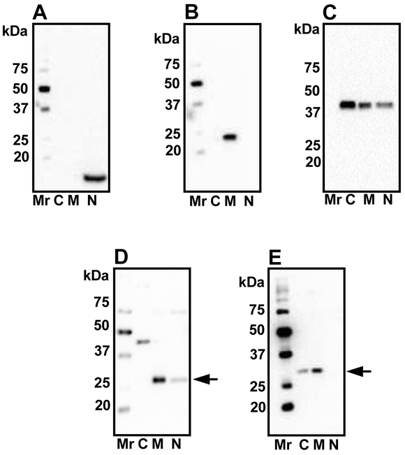
Figure 2: Subcellular localization of SIRT3 and SIRT5.
Mouse lens epithelial cells were cultured, fractionated as described in Material and Methods and immunoblotted using histone H3 antibodies (A), SOD2 (B) and β-actin (C) antibodies. The isolated fractions were Western blotted using SIRT3 (D) and SIRT5 (E) antibodies. Arrows in panels D and E indicate SIRT3 and SIRT5, respectively. C, cytosolic; M, mitochondrial; N, nuclear. Mr, molecular weight markers.

