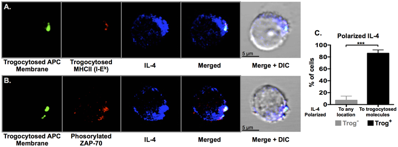
Figure 5. IL-4 is polarized towards trogocytosed molecules and the location of active TCR signaling.
CD4+ T cells were analyzed by confocal microscopy 72-hours after recovery from a standard in vitro trogocytosis assay. Representative optical sections of trog+ cells are shown with (A) trogocytosed APC membrane proteins (green), trogocytosed I-Ek (red), and intracellular IL-4 (blue). (B) Trogocytosed APC membrane proteins (green), phosphorylated-Zap-70 (red) and intracellular IL-4 (blue). Data is representative of five independent experiments, with over 120 individual trog+ CD4+ cells analyzed in total. (C) IL-4+ trog− (grey) and trog+ (black) cells were analyzed for IL-4 polarization, as defined by the localization of intracellular IL-4 to any constrained area for the trog− cells, and polarization towards trogocytosed molecules in the trog+ cells. Error bars represent ±SEM from three independent experiments, *** = p≤ 0.001.

