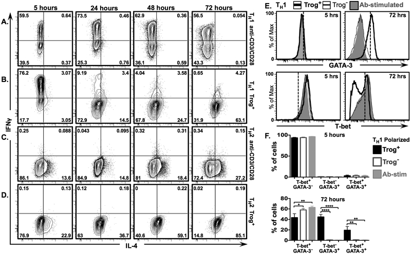
Figure 7. IL-4 expression increases and IFNγ expression decreases in polarized TH1 trog+ cells after removal of APC.
In vitro polarized TH1 and TH2 CD4+ T cells were used in a standard in vitro trogocytosis assay. In parallel, aliquots of polarized cells were stimulated with plate-bound anti-CD3 + anti-CD28 Abs. Fluorescence intensity of IL-4 vs. IFNγ is displayed in representative 2D scatter plots at 5 hrs. (left), 24 hrs., (2nd from left), 48 hrs. (2nd from right), and 72 hrs. (right)for (A) anti-CD3 + anti-CD28 stimulated TH1 polarized cells, (B) TH1 polarized trog+ cells (C) anti-CD3 + anti-CD28 stimulated TH2 polarized, and (D) TH2 polarized trog+ cells. Numbers represent frequency of cells in each quadrant. (E) Representative histograms showing intensity of GATA-3 (top) and T-bet (bottom) expression in TH1 polarized trog+(thick black line) and trog− (thin black line) cells, compared to anti-CD3/CD28 stimulated blasts (shaded grey), at 5 (left), and 72 (right), hours post-recovery. (F) Mean frequency of T-bet+ GATA-3− (left), T-bet− GATA-3+ (middle), and T-bet+ GATA-3+ (right), in vitro polarized TH1 trog+ (black), trog− (grey outline), and anti-CD3/CD28 stimulated cells (shaded grey) at 5 (top), and 72 (bottom) hours post-recovery. Error bars represent ±SEM from three independent experiments, * = p≤ 0.05, ** = p≤ 0.01, *** = p≤ 0.001, and **** = p≤ 0.0001. All data is representative of three independent experiments.

