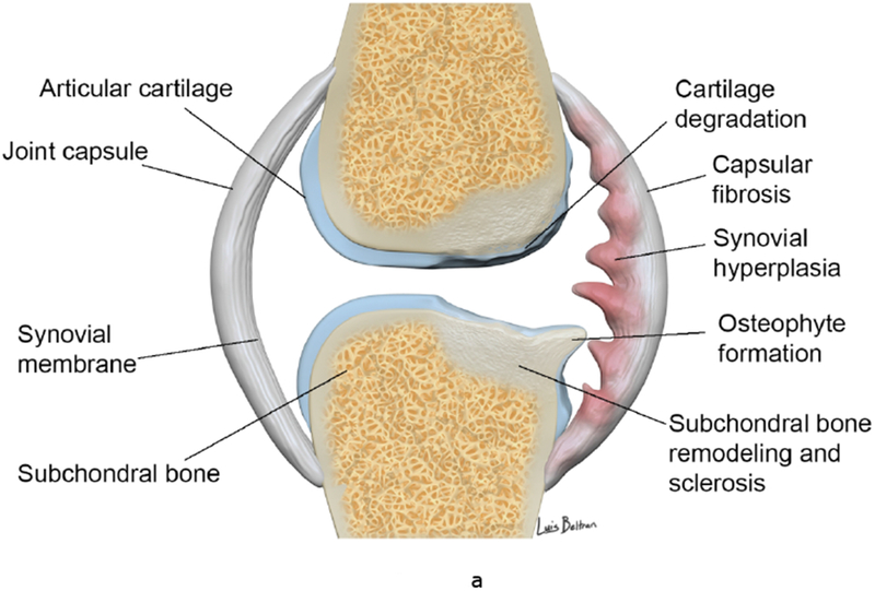
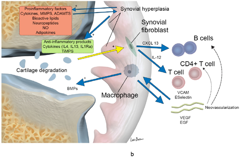
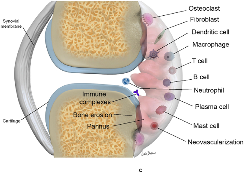
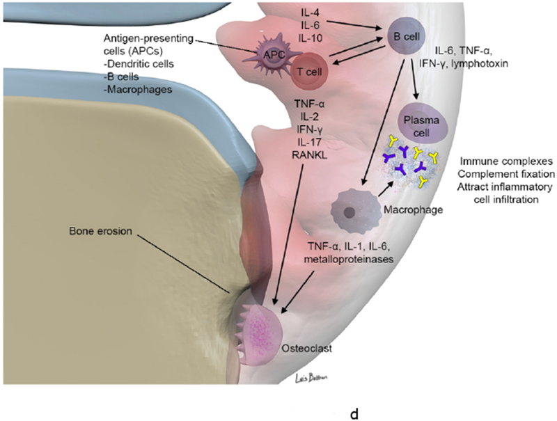
Figure 1.


(a-b). Synovial involvement in OA pathophysiology. Degraded cartilaginous products within synovial fluid are phagocytosed by activated synovial cells within the inflamed synovium which in turn produce proinflammatory and catabolic mediators. Proteolytic enzymes promote cartilage breakdown creating a positive feedback loop. Inflammatory cytokines promoting enzymatic mediators of cartilage catabolism include MMPs and ADAMTS. The inflamed synovium also contributes to osteophyte formation via BMPs. Activated infiltrating macrophages, synovial B cells and T cells amplify this inflammatory response however the synovium and cartilage may also produce counteracting anti-inflammatory products such as IL-4, IL-13, IL1Ra and TiMPS.
(c-d). Synovial involvement in RA pathophysiology. The synovium is infiltrated with mature antigen presenting cells (APCs) (including dendritic cells, macrophages and B cells) and activated T lymphocytes, which interact to initiate and amplify T-cell-dependent immune responses, promoting chronic synovial inflammation. Activated T cells induce secretion of pro-inflammatory cytokines (in particular TNF α) from macrophages and synovial cells in a contact-dependent manner. Macrophages also function as osteoclast precursors, as well as providing specific molecular signals driving osteoclast formation. Numerous osteoclasts are found within the inflamed synovium adjacent to bone, creating resorption pits and causing local bone destruction through specific enzymes and a proton pump that enable them to degrade bone matrix and solubilize calcium. RANKL binds to receptors of pre-osteoclasts and mature osteoclasts and stimulates their activation and differentiation (together with TNF, IL-17 and IL-1). B cells contribute to immune complex formation and complement activation, act as APCs and contribute to T cell activation through expression of costimulatory molecules. B cells both respond to and produce the chemokines and cytokines that promote leukocyte infiltration into the joints, angiogenesis, and synovial hyperplasia.
ADAMTS a disintegrin and metalloproteinase with thrombospondin motifs, BMP bone morphogenetic protein, CXCL13 CXC-chemokine ligand 13, EGF endothelial growth factor, IFN-γ interferon γ, IL interleukin, IL-1Ra IL-1 receptor antagonist, LTB4, leukotriene B4, MMP matrix metalloproteinase, NO nitric oxide, RANKL receptor activator of nuclear factor κB ligand, TiMP tissue inhibitor of metalloproteinase, TNF tumor necrosis factor, VCAM, vascular cell adhesion molecule 1, VEGF vascular endothelial growth factor.
