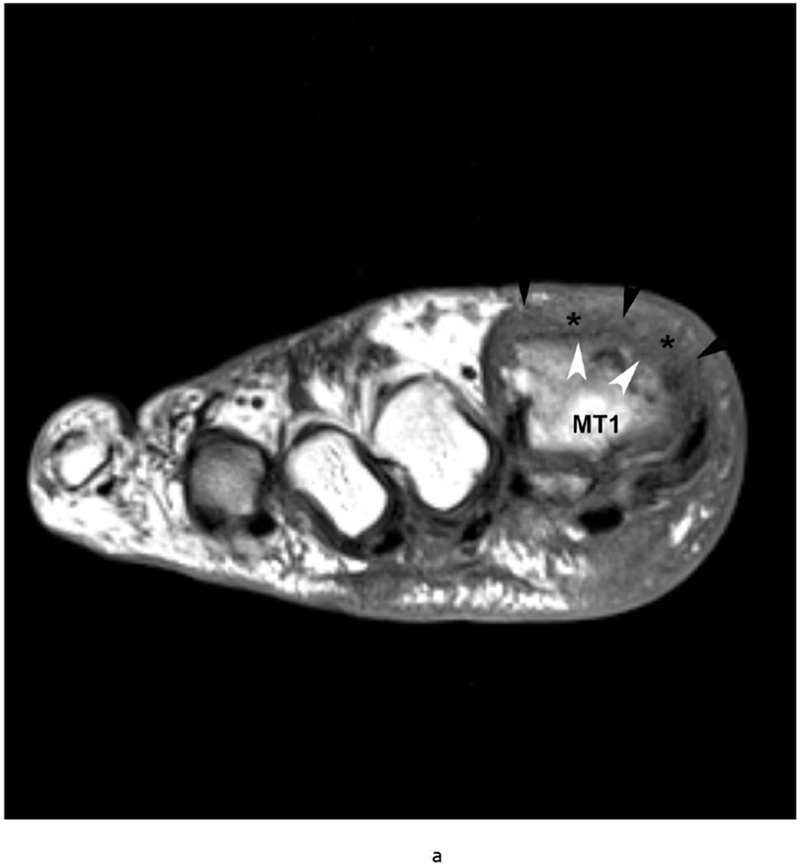
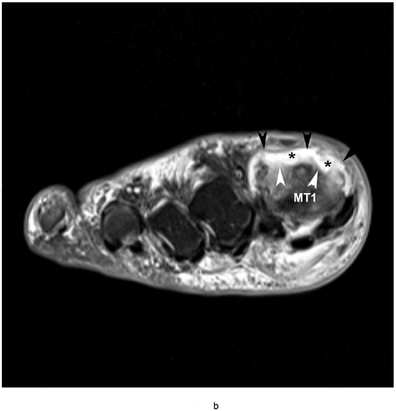
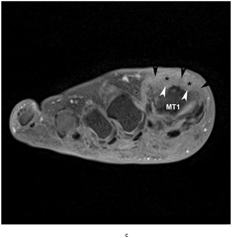
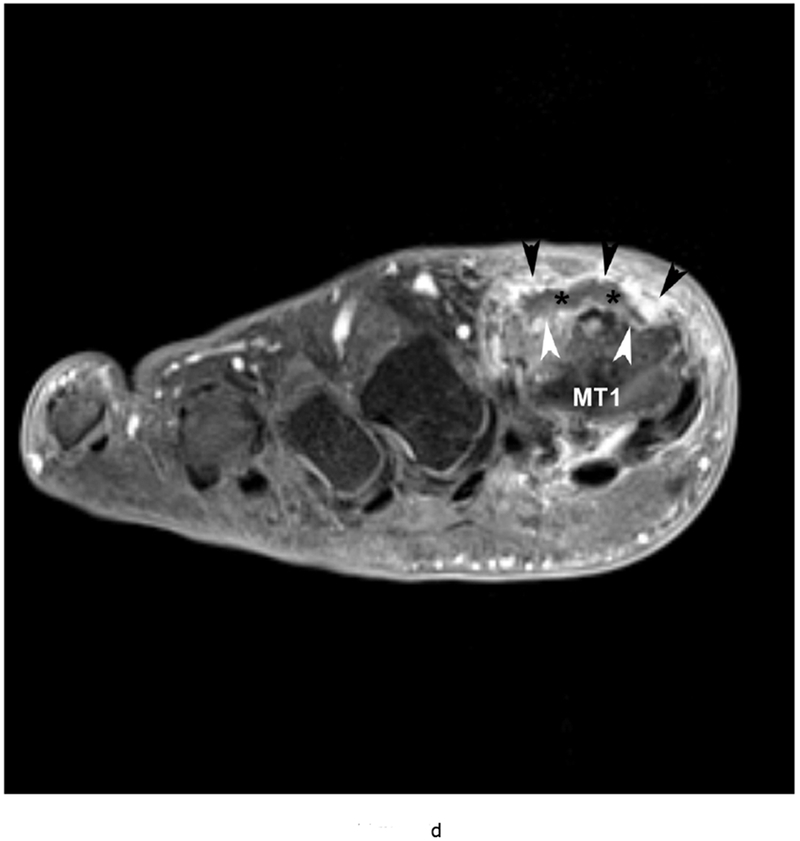
Figure 5.


79 year old male with gout, confirmed by aspiration of monosodium urate (MSU) crystals. Short axis (a) T1 (TR 528 / TE 10) (b) T2 FS (TR 5610/TE 40), (c) T1 FS pre ( TR 612/TE 10) and (d) post Gadovist contrast images (TR 670/TE 12) demonstrating an effusion (*) in association with synovitis of the first metatarsophalangeal joint distending the joint capsule (black arrowheads). There are osseous erosions (white arrowheads) of the first metatarsal head (MT1) with subjacent bone marrow edema.
